neuralib.atlas.brainrender
BrainRender Wrapper
- author:
Yu-Ting Wei
This module provide a CLI-based brainrender wrapper See detail in the https://brainglobe.info/documentation/brainrender/index.html. The wrapper provide three main usage cases, and can be run as command line once the package installed
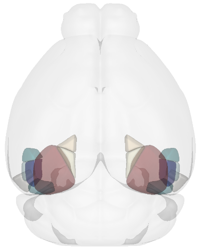
Region reconstruction (area mode)
Plot brain regions
Example of reconstruct the Visual Cortex
brender area -R VISal,VISam,VISl,VISli,VISp,VISpl,VISpm,VISpor --camera top
See the available options use -h option
brender area -h
ROI reconstruction (roi mode)
Plot brain regions with ROIs label
Example of reconstruct ROIs in the Somatosensory Cortex for ipsilateral hemisphere(assume right hemisphere):
brender roi -F <CSV_FILE>
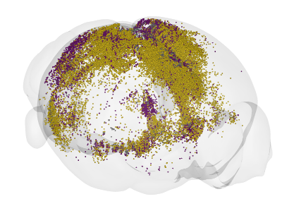
CSV FILE example (auto transformed coordinates space from allen to brainrender):
┌───────────────────────────────────┬─────────┬─────────────┬─────────────┬─────────────┬─────────┬─────────┬────────┬───────────────────────────┬──────────────┬────────┬────────────┬────────────┬────────────┬────────────┬────────────┬───────────┐
│ name                              ┆ acronym ┆ AP_location ┆ DV_location ┆ ML_location ┆ avIndex ┆ channel ┆ source ┆ abbr                      ┆ acronym_abbr ┆ hemi.  ┆ merge_ac_0 ┆ merge_ac_1 ┆ merge_ac_2 ┆ merge_ac_3 ┆ merge_ac_4 ┆ family    │
│ ---                               ┆ ---     ┆ ---         ┆ ---         ┆ ---         ┆ ---     ┆ ---     ┆ ---    ┆ ---                       ┆ ---          ┆ ---    ┆ ---        ┆ ---        ┆ ---        ┆ ---        ┆ ---        ┆ ---       │
│ str                               ┆ str     ┆ f64         ┆ f64         ┆ f64         ┆ i64     ┆ str     ┆ str    ┆ str                       ┆ str          ┆ str    ┆ str        ┆ str        ┆ str        ┆ str        ┆ str        ┆ str       │
╞═══════════════════════════════════╪═════════╪═════════════╪═════════════╪═════════════╪═════════╪═════════╪════════╪═══════════════════════════╪══════════════╪════════╪════════════╪════════════╪════════════╪════════════╪════════════╪═══════════╡
│ Ectorhinal area/Layer 5           ┆ ECT5    ┆ -3.03       ┆ 4.34        ┆ -4.5        ┆ 377     ┆ gfp     ┆ aRSC   ┆ Ectorhinal area           ┆ ECT          ┆ contra ┆ ECT        ┆ ECT        ┆ ECT        ┆ ECT        ┆ ECT        ┆ ISOCORTEX │
│ Perirhinal area layer 6a          ┆ PERI6a  ┆ -3.03       ┆ 4.42        ┆ -4.37       ┆ 372     ┆ gfp     ┆ aRSC   ┆ Perirhinal area           ┆ PERI         ┆ contra ┆ PERI       ┆ PERI       ┆ PERI       ┆ PERI       ┆ PERI       ┆ ISOCORTEX │
│ …                                 ┆ …       ┆ …           ┆ …           ┆ …           ┆ …       ┆ …       ┆ …      ┆ …                         ┆ …            ┆ …      ┆ …          ┆ …          ┆ …          ┆ …          ┆ …          ┆ …         │
│ Ventral auditory area layer 6a    ┆ AUDv6a  ┆ -2.91       ┆ 3.52        ┆ 4.46        ┆ 156     ┆ rfp     ┆ pRSC   ┆ Ventral auditory area     ┆ AUDv         ┆ ipsi   ┆ AUD        ┆ AUD        ┆ AUD        ┆ AUD        ┆ AUDv       ┆ ISOCORTEX │
│ Ectorhinal area/Layer 6a          ┆ ECT6a   ┆ -2.91       ┆ 4.14        ┆ 4.47        ┆ 378     ┆ rfp     ┆ pRSC   ┆ Ectorhinal area           ┆ ECT          ┆ ipsi   ┆ ECT        ┆ ECT        ┆ ECT        ┆ ECT        ┆ ECT        ┆ ISOCORTEX │
│ Temporal association areas layer… ┆ TEa5    ┆ -2.91       ┆ 4.02        ┆ 4.55        ┆ 365     ┆ rfp     ┆ pRSC   ┆ Temporal association area ┆ TEa          ┆ ipsi   ┆ TEa        ┆ TEa        ┆ TEa        ┆ TEa        ┆ TEa        ┆ ISOCORTEX │
└───────────────────────────────────┴─────────┴─────────────┴─────────────┴─────────────┴─────────┴─────────┴────────┴───────────────────────────┴──────────────┴────────┴────────────┴────────────┴────────────┴────────────┴────────────┴───────────┘
See how to create the csv after ccf pipeline
from neuralib.atlas.ccf.classifier import RoiClassifier
from neuralib.atlas.ccf.core import AbstractCCFDir
root = ...
ccf_dir = AbstractCCFDir(root, with_overlap_sources=False)
classifier = RoiClassifier(ccf_dir, plane='coronal')
df = classifier.parsed_df
Example ccf data folder structure in (AbstractCCFDir())
See the available options use -h option
brender roi -h
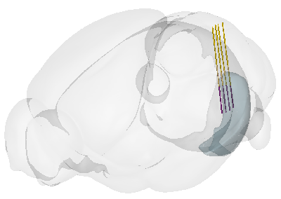
Probe reconstruction (probe mode)
Reconstruct the probe (or shanks) in accordance with trajectory labeling (e.g., DiI, DiO or lesion track…)
Prepare CSV file from ccf pipeline:
┌───────────────────────────────────┬─────────┬─────────────┬─────────────┬─────────────┬─────────┐
│ name                              ┆ acronym ┆ AP_location ┆ DV_location ┆ ML_location ┆ avIndex │
│ ---                               ┆ ---     ┆ ---         ┆ ---         ┆ ---         ┆ ---     │
│ str                               ┆ str     ┆ f64         ┆ f64         ┆ f64         ┆ i64     │
╞═══════════════════════════════════╪═════════╪═════════════╪═════════════╪═════════════╪═════════╡
│ Primary visual area layer 6a      ┆ VISp6a  ┆ -3.81       ┆ 1.92        ┆ -3.12       ┆ 191     │
│ optic radiation                   ┆ or      ┆ -4.08       ┆ 2.33        ┆ -3.12       ┆ 1217    │
│ Posterolateral visual area layer… ┆ VISpl6a ┆ -4.28       ┆ 2.29        ┆ -3.12       ┆ 198     │
│ Posterolateral visual area layer… ┆ VISpl5  ┆ -4.52       ┆ 2.17        ┆ -3.12       ┆ 197     │
│ Subiculum                         ┆ SUB     ┆ -3.93       ┆ 4.36        ┆ -3.3        ┆ 536     │
│ Entorhinal area medial part dors… ┆ ENTm5   ┆ -4.19       ┆ 4.39        ┆ -3.3        ┆ 515     │
│ Entorhinal area medial part dors… ┆ ENTm2   ┆ -4.44       ┆ 4.39        ┆ -3.3        ┆ 510     │
│ Entorhinal area medial part dors… ┆ ENTm1   ┆ -4.66       ┆ 4.29        ┆ -3.3        ┆ 509     │
└───────────────────────────────────┴─────────┴─────────────┴─────────────┴─────────────┴─────────┘
Row number equal to shank numbers * 2 (in the example, use the 4 shanks NeuroPixel probe), 2 points indicate the most dorsal & ventral detected signals on the serial brain slices
Use
-Pto specify the slice cutting orientation {coronal,sagittal,transverse}. If multiple shanks were inserted along the AP axis, assume do the sagittal plane, if inserted along the ML axis, then assume the coronal planeAssume shanks are not bended to perform interpolation based on the insert depth
-D
Example of Above csv file for targeting the left Entorhinal cortex (ENT) using 4 shanks NeuroPixel probe:
brender probe -F <CSV_FILE> -D 3000 -P sagittal -R ENT -H left
See the available options use -h option
brender probe -h